
When a suspicious site is detected in your breast through a breast self-exam or on a screening mammogram, your doctor may request an ultrasound of the breast tissue. A breast ultrasound is a scan that uses penetrating sound waves that do not affect or damage the tissue and cannot be heard by humans. The breast tissue deflects these waves causing echoes, which a computer uses to paint a picture of what’s going on inside the breast tissue. A mass filled with liquid shows up differently than a solid mass.
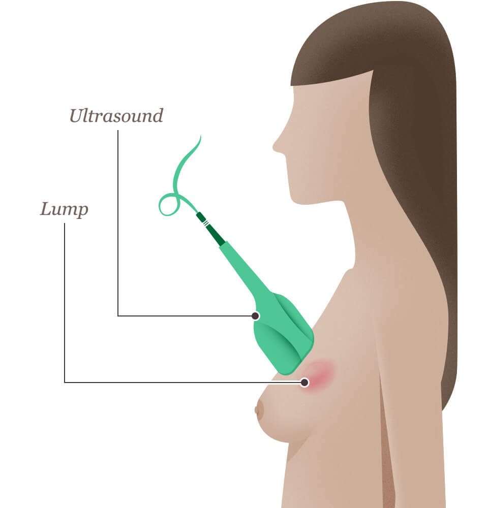
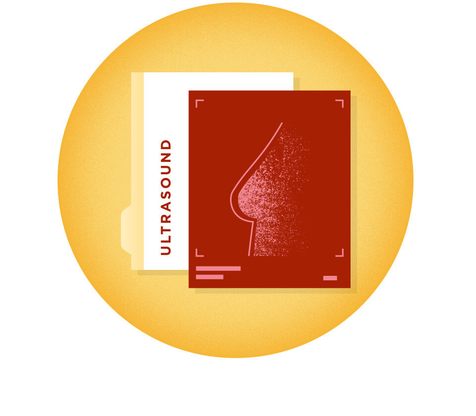
Ultrasound Results: Breast Sonogram
The detailed picture generated by the ultrasound is called a “sonogram.” Ultrasounds are helpful when a lump is large enough to be easily felt, and the images can be used to further evaluate the abnormality.
A breast ultrasound can provide evidence about whether the lump is a solid mass, a cyst filled with fluid, or a combination of the two. While cysts are typically not cancerous in most cases, a solid lump may be a cancerous tumor. Healthcare professionals also use this diagnostic method to help measure the exact size and location of the lump and get a closer look at the surrounding tissue like the lymph nodes in the axilla (armpit).
